Magnetic Resonance Imaging (MRI) has revolutionized the way doctors assess neurological conditions. By creating detailed images of the brain and spinal cord, MRI helps detect abnormalities that indicate neurological disorders. Here is more information on how it works, its role in identifying structural changes, and how it contributes to understanding brain function in neurological evaluations:
How MRI Works
MRI scans use powerful magnetic fields and radio waves to create high-resolution images of the body’s internal structures. Unlike X-rays or CT scans, it does not involve radiation, making it a widely used tool in brain imaging. During a scan, the patient lies inside a large, tube-like machine. The magnetic field causes protons in body tissues to align. Radio waves then disrupt this alignment, emitting signals that the machine converts into detailed images.
These images offer unparalleled clarity of soft tissues, which makes MRI highly effective for studying the brain and spinal cord. Specialized techniques, such as contrast enhancement or diffusion-weighted imaging. This allows doctors to highlight specific abnormalities or track the movement of water molecules in brain tissues. This level of detail is key for diagnosing conditions like tumors, strokes, or multiple sclerosis.
Which Structural Issues Get Detected
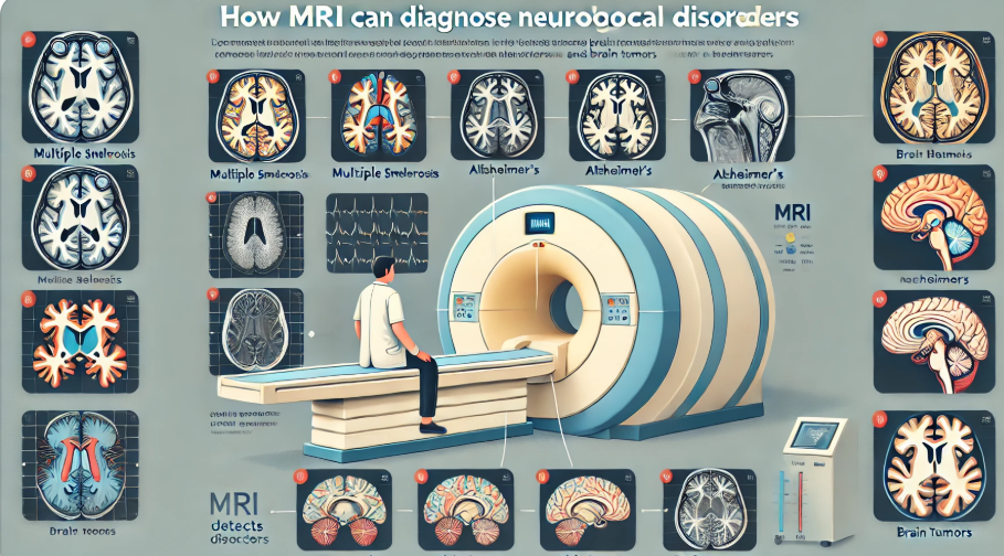
MRI is particularly useful in identifying structural abnormalities that may signal neurological disorders. Conditions such as tumors, multiple sclerosis, and traumatic brain injuries often cause physical changes that MRI can detect with precision. This makes it a necessary tool for early diagnosis and treatment planning.
In multiple sclerosis, MRI can reveal lesions or plaques in the brain and spinal cord caused by the degradation of the protective covering around nerve fibers. These lesions would be difficult to identify with other imaging techniques. In cases of brain tumors, it displays the location, size, and impact of the tumor on surrounding tissues, guiding both diagnosis and treatment planning.
MRI can also provide clarity about the extent of damage in cases of traumatic brain injuries. It helps visualize any swelling, bleeding, or changes in the brain’s structure that could explain symptoms like memory loss or impaired motor functions. This detailed imaging is key for doctors to develop effective treatment plans.
How MRI Evaluates Brain Function
Beyond structural imaging, this technology can also assess brain activity and connectivity through functional MRI (fMRI). This specialized application measures changes in blood flow in the brain, indicating regions that are active during specific tasks. This information proves particularly useful in conditions such as epilepsy. They can identify functional abnormalities in brain regions responsible for seizures, assisting in diagnosis and treatment approaches.
For neurodegenerative conditions like Alzheimer’s disease, it aids in understanding how parts of the brain may shrink or lose function over time. Functional MRI is also instrumental in pre-surgical planning for patients undergoing brain surgery. It helps surgeons minimize potential risks by mapping key areas, such as those responsible for speech or motor skills.
Find Diagnostic Imaging Services
MRI has become an indispensable tool in diagnosing neurological disorders. It provides unmatched detail in imaging the brain’s structure and function, making it necessary for evaluating certain conditions. By leveraging MRI, healthcare professionals gain valuable insights for accurate diagnoses and effective treatment planning.
- Integrating IV Therapy With Local Health Retreats for an Enhanced Wellness Experience in Temecula
- Recognizing the Symptoms of a Rotator Cuff Tear
- Tips for Managing Pain Post-Gallbladder Surgery
- How to Prepare for Your First Visit to a Dermatologist
- How a Pain Management Doctor Can Help Improve Your Quality of Life
Leave a Reply