Accurate diagnosis is key to managing orthopedic conditions. Advances in medical imaging allow physicians to see the body like never before. Body imaging techniques like X-rays, CT scans, MRIs, and ultrasounds identify issues in muscles, bones, and joints. These methods make orthopedic assessments more reliable and efficient. Understanding these technologies helps patients better understand their diagnoses.
What Is Body Imaging?
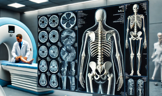
Orthopedic body imaging refers to medical techniques and technologies used to visualize bones, muscles, and joints. These methods produce detailed images of the internal structures of the body, aiding physicians in identifying specific issues. Imaging is pivotal in modern medicine, offering a non-invasive approach to assess various conditions.
X-rays are one of the most commonly used imaging methods. They work by using electromagnetic radiation to capture images of dense structures, like bones. CT scans combine X-ray technology with computer processing to provide cross-sectional images of the body.
MRI uses a magnetic field and radio waves to generate highly detailed images, particularly of soft tissues like ligaments and cartilage. Ultrasound creates real-time images using sound waves, making it especially useful for muscles and blood vessels. These tools allow orthopedic professionals to study the internal body without surgical exploration, offering a safer and more efficient diagnostic option.
How Are These Techniques Used for Diagnosing Orthopedic Conditions?
Body imaging is used to detect abnormalities, monitoring progress, and evaluating treatment effectiveness. Each type of imaging serves unique purposes, offering specific advantages based on the condition being assessed. X-rays are a quick and reliable tool for diagnosing fractures and bone misalignments, such as broken arms or legs. CT scans provide a more detailed view for complex bone injuries, small fractures, and joint structures, aiding in precise treatment planning.
For soft tissue evaluations, MRI is particularly effective in diagnosing ligament tears, cartilage damage, or spinal disc problems. Its ability to provide detailed images of both bones and surrounding tissues makes it invaluable for identifying issues other methods might miss. Ultrasound excels at assessing muscle strains, tendon injuries, and inflammation in joints. It also plays a key role in visualizing blood flow, which is especially useful in cardiovascular evaluations tied to orthopedic care.
Imaging tools provide healthcare providers with detailed insights, enabling precise diagnoses of various conditions. This level of clarity helps provide an accurate understanding of the issue at hand. With this information, personalized and targeted treatment plans can be developed to address each patient’s specific needs effectively.
What Conditions Can These Techniques Discover?
Orthopedic imaging techniques are highly versatile, aiding in the detection and analysis of numerous conditions across the musculoskeletal, abdominal, and cardiovascular systems. For musculoskeletal issues, X-rays are commonly used to identify fractures, dislocations, or bone deformities. MRIs are used to diagnose injuries such as rotator cuff tears or knee ligament damage. CT scans, on the other hand, are often employed to evaluate complex fractures or spinal problems that may require surgical intervention.
Beyond musculoskeletal conditions, imaging plays a role in detecting abdominal and pelvic issues that impact orthopedic health. CT scans can identify hernias or pelvic abnormalities that affect joint function. Ultrasounds are particularly effective in spotting soft tissue inflammation or vascular issues that cause pain or restricted mobility. These insights make imaging a foundational tool for addressing interconnected health concerns that may not seem orthopedic at first glance.
Cardiovascular imaging also has applications in orthopedic care by evaluating circulatory issues that may influence joint health. Restricted blood flow can lead to joint pain or swelling, and ultrasound provides real-time visualization of blood flow to aid in diagnosis. With its ability to assess a wide range of conditions, imaging is an integral component of comprehensive medical evaluations in orthopedics and beyond.
Precision in Diagnosis Begins Here
Advances in body imaging have transformed how orthopedic conditions are detected and treated. Techniques like X-rays, CT scans, MRI, and ultrasound provide invaluable information about musculoskeletal, abdominal, and cardiovascular systems. These methods continue to enhance the precision and efficiency of orthopedic care. If you’re seeking expert guidance for orthopedic assessments or treatment, reach out to your healthcare provider today. Understanding your condition starts with accurate diagnostics, and imaging plays a central role in that process.
- Personalized Approaches in Therapy Services for Lasting Change
- How to Choose the Right Cardiologist for Your Heart Care
- What To Expect During Your First Appointment With an Orthopedic Surgeon
- Understanding the Various Surgical Weight Loss Options Available
- Understanding Common Injuries Treated by Sports Medicine Specialists
Leave a Reply